Brainwashing Tumours

I’ve been ever so lucky in spending a lifetime in science — particularly in cancer biology because, even in the 21st century, every couple of weeks or so you spot some piece of work that makes your jaw drop as you mumble “Wow! That’s stunning”. It’s true that nowadays the breathtaking bit often comes from the staggering panoply of techniques that have been thrown at a problem and the vast amounts of data presented.

But once in a while a “Wow” moment turns up because some inquisitive soul has peered into a nook that no one has bothered looking at before and found something amazing. Something of which almost everyone in the field would have said “Nah! Forget it! Can’t happen!! You’re wasting your time!!!”

We talked about a recent example back in March: the finding that bugs prefer to infect cancer cells rather than their normal bretheren (remarkable in itself) and that, when they do so, they can help tumours to spread — i.e. metastasize (Mapping the bug army).

Now comes an even more startling and unpredicted finding and it’s about a type of primary brain tumour — malignant gliomas. One of the most common categories of primary brain tumour, glioma is an umbrella term for cancers of the glial cells that surround nerve endings in the brain. Glioblastoma is a type of glioma. Treatment is by surgery, radiotherapy and chemotherapy but the 5-year survival rate remains at around 5% and its progression is usually accompanied by progressive cognitive decline. How tumours disrupt brain function is not understood but possible mechanisms are summarised below (a-d). Broadly speaking these are the rather obvious ones of tumours invading or compressing tissues or recruiting blood supplies form the host.

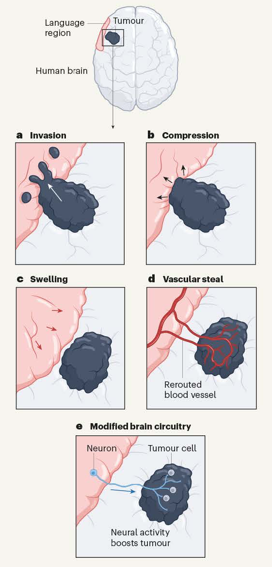
Ways in which brain tumours might affect brain activity. These include (a) invasion, (b) compression, (c) swelling and (d) recruiting a blood supply (‘vascular steal’). The recent evidence from Krishna et al. reveals that tumours can form connections (synapses) with neurons. When the neurons are active (e.g., engaged in a language task) tumour growth is boosted. From Ibrahim and Taylor 2023.

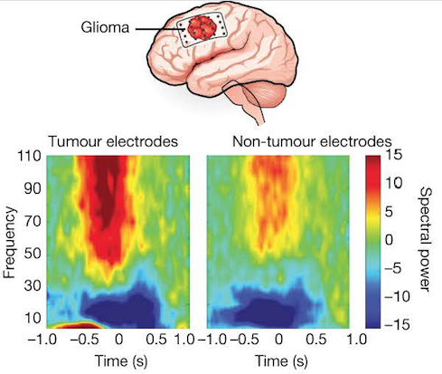
High-grade gliomas remodel long-range functional neural circuits. Top: Scheme showing a dominant hemisphere glioblastoma (i.e. a tumour in the left hemisphere — the one that controls the dominant right hand). Lower panels: Electrocorticography (a type of electrophysiological monitoring that uses electrodes placed directly on the exposed surface of the brain to record electrical activity from the cerebral cortex). The spectral data show clear separation of frequencies across tumour and non-tumour electrodes. From Krishna et al., 2023.

Some thought!

The electrical activity shown in the above figure was recorded in individuals undergoing brain surgery who were asked to name items in pictures whilst their brain-surface activity was recorded. The data show that activity in regions of the brain involved in a language task also drove activity in tumour-associated regions that do not normally function in language processing. High functional connectivity associated with neuronal signalling in tumours predicts aggressive tumour behaviour, cognitive decline and poor survival.

Unsurprisingly, functional connectivity varied across tumours and regions were classified as having high or low functional connectivity (HFC or LFC). HFC regions had raised levels of thrombospondin 1, an adhesive glycoprotein that mediates cell-to-cell and cell-to-matrix interactions — it’s involved in synapse formation and is normally secreted by astrocytes.

Moreover, addition of thrombospondin 1 to LFC regions of gliomas co-cultured with neurons induced behaviour resembling that of co-cultures with cells from the HFC region of the tumour. Conversely, when the TSP-1 inhibitor gabapentin was added to the co-cultures, glioma proliferation was reduced. Furthermore, glioma-infiltrated brain tissue enriched in TSP-1 formed synapses when grafted into the hippocampal region of the mouse brain in vivo.

Astounding!

It’s been known for a while that working synapses can link neurons to gliomas. The latest, extraordinary work from Saritha Krishna and colleagues in San Francisco has shown that conscious thought, as manifested by speech, can actually promote tumour growth. Astonishingly, high-grade gliomas functionally remodel neural circuits in the human brain, which both promotes tumour progression and impairs cognition. Not only a staggering thought but also food for thought in that the thrombospondin data clearly point the way to desperately needed new therapeutic approaches for these cancers.

References

Ibrahim GM, Taylor MD. How thought itself can drive tumour growth. Nature. 2023 May;617(7961):469-471. doi: 10.1038/d41586-023-01387-1. PMID: 37138055.

Krishna, S., Choudhury, A., Keough, M.B. et al. Glioblastoma remodelling of human neural circuits decreases survival. Nature 617, 599–607 (2023). https://doi.org/10.1038/s41586-023-06036-1

I Know What I Like


I guess most of us at some time or other will have stood gazing at a painting for a while before muttering ‘Wow, that’s awesome’ or words to that effect if we’re not into the modern argot. Some combination of subject, style and colour has turned our crank and left us thinking we wouldn’t mind having that on our kitchen wall.

Given the thousands of years of man’s daubing and the zillions of forms that have appeared from pre-historic cave paintings through Eastern painting, the Italian Renaissance, Impressionism, Dadaism and the rest to Pop Art, it’s amazing that everyone isn’t a fanatic for one sort or another. The sane might say the field’s given itself a bad name by passing off tins of baked beans, stuff thrown at a canvas and unmade beds as ‘art’ but, even so, it seems odd that it remains a minority obsession.

Can science help?

Science is wonderful, as we all know, but the notion that it might arouse the collective artistic lust seems fanciful. Nevertheless, unnoticed by practically everyone, our vast smorgasbord of smears has been surreptitiously joined over the last 30 years by a new form: an ever-expanding avalanche of pics created by biologists trying to pin down how animals work at the molecular level. The crucial technical development has been the application of fluorescence in the life sciences: flags that glow when you shine light on them and can be stuck on to molecules to track what goes on in cells and tissues. The pioneer of this field was Roger Tsien who died, aged 64, in 2016.

Because this has totally transformed cell biology we’ve run into lots of brilliant examples in these pages — recently in Shifting the Genetic Furniture, in Caveat Emptor and John Sulston: Biologist, Geneticist and Guardian of our Heritage and in the use of red and green tags for picking out individual types of proteins that mark mini-cells within cells in Lorenzo’s Oil for Nervous Breakdowns.

To mark the New Year this piece looks at science from a different angle by focussing not on the scientific story but on the beauty that has become a by-product of this pursuit of knowledge.

Step this way: entrance free

So let’s take a stroll through our science gallery and gaze at just a few, randomly selected works of art.

  1. Cells grown in culture:

This was one of the first experiments in my laboratory using fluorescently labelled antibodies, carried out by a student, Emily Hayes, so long ago that she now has a Ph.D., a husband and two children. The cells are endothelial cells (that line blood vessels). Blue: nuclei; green: F-actin; red: Von Willebrand factor, a protein marker for endothelium.


  1. Two very recent images taken by my colleague Roderik Kortlever of a senescent mouse fibroblast and of mouse breast tissue:


3. Waves of calcium in firing neurons:

One of my fondest memories is helping to do the first experiment that measured the level of calcium within a cell, carried out with my colleague the late Roger Tsien and two other friends. I only grew the cells: Roger had designed and made the molecule, quin2. We didn’t know it at the time but Roger’s wonder molecule was the first of many intracellular ‘reporters.’ Roger shared the 2008 Nobel Prize in Chemistry for his discovery and development of the green fluorescent protein with organic chemist Osamu Shimomura and neurobiologist Martin Chalfie.

This wonderful video of a descendant of quin2 in nerve cells was made in Dr. Sakaguchi’s lab at Iowa State University.


4. Calcium wave flooding a fertilized egg: Taro Kaneuchi and colleagues at the Tokyo Metropolitan University:

Click for a time-lapse movie of an egg cell that has been artificially stimulated to show the kind of calcium change that happens at fertilization. In this time-lapse movie the calcium level reaches a maximum signal intensity after about 30 min before gradually decreasing to the basal level.


5. The restless cell (1):

This movie shows how protein filaments in cells can continuously break down and reform – called treadmilling. Visualised in HeLa cells using a green fluorescent protein that sticks to microtubules (tubular polymers made up of the protein tubulin) by HAMAMATSU PHOTONICS.


6. The restless cell (2):

This movie shows how mitochondria (organelles within the cell) are continuously changing shape and moving within the cell’s interior (cytosol). Red marks the mitochondria; green DNA within the nucleus. HAMAMATSU PHOTONICS.


7. Cell division:

Pig kidney cells undergoing mitosis. Red marks DNA (nucleus); green is tubulin: HAMAMATSU PHOTONICS.


8. DNA portrait of Sir John Sulston by Marc Quinn commissioned by the National Portrait Gallery:This image looks a bit drab in the present context but in some ways it’s the most dramatic of all. John Sulston shared the 2002 Nobel Prize in Physiology or Medicine with Sydney Brenner and Robert Horvitz for working out the cell lineage of the roundworm Caenorhabditis elegans (i.e. how it develops from a single, fertilized egg to an adult). He went on to sequence the entire DNA of C. elegans. Published in 1998, it was the first complete genome sequence of an animal — an important proof-of-principle for the Human Genome Project that followed and for which Sulston directed the British contribution at the Sanger Centre in Cambridgeshire, England. The project was completed in 2003.

The portrait shows colonies of bacteria in a jelly that, together, carry all Sulston’s DNA. This represents DNA cloning in which DNA fragments, taken up by bacteria after insertion into a circular piece of DNA (a plasmid), are multiplied to give many identical copies for sequencing.


9. “Brainbow” mice by Tamily Weissman at Harvard University:

The science behind this astonishing image builds on the work of Roger Tsien. Mice are genetically engineered to carry three different fluorescent proteins corresponding to the primary colours red, yellow and blue. Within each cell recombination occurs randomly, giving rise to different colours. The principle of mixing primary colours is the same as used in colour televisions.  In this view individual neurons in the brain (specifically a layer of the hippocampus) project their dendrites into the outer layer. Other magnificent pictures can be seen in the Cell Picture Show.

It’s certainly science – but is it art?

A few years ago the Fitzwilliam Museum in Cambridge staged Vermeer’s Women, an exhibition of key works by Johannes Vermeer and over thirty other masterpieces from the Dutch ‘Golden Age’. I tried the experiment of standing in the middle of each room and picking out the one painting that, from a distance, most caught my amateur eye. Funny thing was: not one turned out to be by the eponymous star of the show! Wondrous though Vermeer’s paintings were, the ones that really took my fancy were by Pieter de Hooch, Samuel van Hoogstraten and Nicolaes Maes, guys I’d never heard of.

Which made the point that you don’t need to be a big cheese to make a splash and that in the new Dutch Republic of the 17th century, the most prosperous nation in Europe, there was enough money to keep a small army of splodgers in palettes and paint. Skillful and incredibly patient though these chaps were, they simply used the tools available to paint what they saw in the world before them — as for the most part have artists down the ages.

But hang on! Isn’t that what we’ve just been on about? Scientists applying enormous skill and patience in using the tools they’ve developed to visualize life — to image what Nature lays before them. So the only difference between the considerable army of biological scientists around the world making a new art form and the Old Masters is that the newcomers are unveiling life — as opposed to the immortalizing a rather dopy-looking aristocrat learning to play the virginal or some-such.

Controversial?

Not really. Let’s leave the last word to Roger Tsien. In our final picture there are eight bacterial colonies each expressing a different colour of fluorescent protein arranged to grow as a San Diego beach scene in a Petri dish. It became the logo of Roger’s laboratory.