First Responders

What better way to celebrate a New Year than with a visit to an art gallery? Well, an alternative might be to scan the cell biology literature but, if you want to cut corners, revisit some of the recent items in this blog.

In Boldly Going we described a method for mapping cell types within tissues on the basis of RNA expression. This identified thousands of RNAs in each cell and, in picking out their spatial pattern, produced images that resemble a pointillist painting — hence the package was christened Seurat by its creators.

That’s very informative but the cellular crock of gold, so to speak, is protein expression — because they do the work in cells — so the next step (Mosaic Masterpieces) revealed how a panel of antibodies could pick out 36 different types of cell in the environment of breast tumours and how the patterns of cells related to patient survival. The tagged cell images also resemble paintings using the pointillism technique developed by Georges Seurat and Paul Signac towards the end of the 19th century, a comparison strengthened by the remarkable comment said to have been made by Seurat that “Great things are done by a series of small things brought together. Some say they see poetry in my paintings; I see only science.”

The next small thing

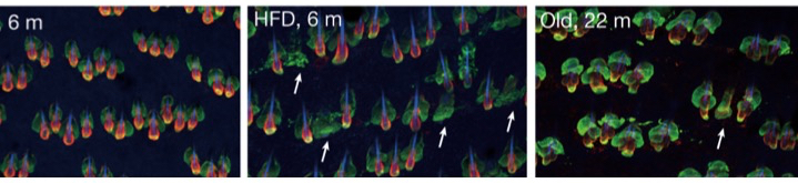
Xiao Qian Wang, Raza Ali and colleagues from the CRUK Cambridge Institute and numerous centres across Europe and extending to Taiwan have given a perfect example of the truism of Seurat’s words in contemporary science. Taking up the methodology of Mosaic Masterpieces, the question they asked was: “Why does immune checkpoint blockade (ICB) work in some patients with triple-negative breast cancer but not in others?” [ICB uses a class of drugs known as immune checkpoint inhibitors {e.g., pembrolizumab (Keytruda) and ipilimumab (Yervoy)}. Much like other immunotherapies, the goal of checkpoint blockade immunotherapy is to strengthen the body’s immune system and enhance its ability to fight off harmful invaders]. Because ICB targets cell-cell interactions, they looked at the type of cells in the tumour microenvironment and how it is changed by ICB (it’s an extension of the work of Keren & Co on triple negative breast cancer described in Mosaic Masterpieces). They tracked 43 different proteins and the montage below gives an artistic impression of their efforts.

Representative images of protein expression in tumour samples. 43 different proteins were detected by imaging mass cytometry (IMC). The image labels refer to proteins detected on specific cells. E.g., CD20 is a general marker for most B-cells, TOX and PD-1 are cytotoxic T cells, TOX and OX40 are T helper cells. PanCK = cytokeratins (keratins).To pursue any of these proteins try The Human Gene Nomenclature database at: https://www.genenames.org/tools/search/.White scale bars = 50 μm. From Wang et al., 2023.

Never mind the art, what about the science?

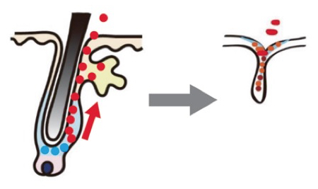
The critical findings were that the levels of a sub-population of T cells (CD8+TCF1+T cells) and of tumour cells that express MHCII+ were dominant predictors of response. In addition, interactions between tumour cells and two other types of immune cells (B cells and granzyme B+ T cells) were important. In other words, ICB treatment remodels the tumour environment and tumours that respond to treatment have lots of granzyme B+ T cells whereas resistant tumours are rich in CD15+ cancer cells. This effect is shown schematically in the image below. [Terminology: CD8 (cluster of differentiation 8): a transmembrane glycoprotein that is a co-receptor for the T-cell receptor (TCR). TCF1 is a transcription factor that is essential for early T cell development. Granzyme B is a caspase-like serine protease that is released by cytotoxic lymphocytes to kill virus-infected and tumour cells. Major histocompatibility complex (MHC) class II molecules present processed antigens, derived primarily from exogenous sources, to CD4(+) T-lymphocytes].

Spatial interactions in a responsive tumour. Here immunotherapy has given rise to a high level of epithelial–CD8+GZMB+T cell interactions. White lines mark tumour epithelial cells interacting with CD8+GZMB+T cells. White scale bar = 50 μm. From Wang et al., 2023.

Taken together these results show that the organization of cell types within the tumour microenvironment determines whether ICB works or not. The key implication is that biopsies taken in the early stages of ICB may give a clear guide as to whether the treatment is working.

As Seurat might have observed, ‘Small steps, small steps’. Very true — but this work tags over 40 different types and sub-types of cell coming and going in the tumour locale. We can be sure that none of them are there as a contribution to art. Thus we need to tease apart all these players in the tumour ecosystem if we are to reach the goal of precision immuno-oncology.

Reference

Wang XQ, Danenberg E, Huang CS, Egle D, Callari M, Bermejo B, Dugo M, Zamagni C, Thill M, Anton A, Zambelli S, Russo S, Ciruelos EM, Greil R, Győrffy B, Semiglazov V, Colleoni M, Kelly CM, Mariani G, Del Mastro L, Biasi O, Seitz RS, Valagussa P, Viale G, Gianni L, Bianchini G, Ali HR. Spatial predictors of immunotherapy response in triple-negative breast cancer. Nature. 2023 Sep;621(7980):868-876. doi: 10.1038/s41586-023-06498-3. Epub 2023 Sep 6. PMID: 37674077; PMCID: PMC10533410.

Less On Top Of More

Many moons ago powdered wigs were all the rage — a fashion seemingly started by The Sun King, aka Louis XIV, who noticed he was going bald at the tender age of 17 and no doubt felt this might limit his sex appeal. Mind you, as syphilis is thought to have caused his hair failure, you might think that quite enough of a turn-off — though maybe not in the jolly old Court of Versailles.

Whatever, when Charles II followed the trend in England, it was wigs for all.

Shifting an itch

In fact there was some science behind the fad in that, back in the 17th century, head lice (nits as we might say) were a problem. When heads were shaved and wigs fitted the nits simply moved upstairs and could be dealt with on laundry day.

What’s new?

Wigs have come and gone but man’s hair anxiety remains — as judged by the number of hairy potions you can buy on line and the fact that the U.S. Food and Drug Administration has approved two drugs to treat male pattern baldness (i.e. a receding front hair line or a bald spot). You can even buy a baseball cap with built-in hair re-growth promoting lasers for £1200.

There’s nothing odd about hair loss — it happens all the time to the tune of up to 100 hairs a day — and pattern hair loss is also common, appearing (disappearing might be a better word) in about 50% of men and a quarter of women by age 50 — generally put down to genetics, age and male hormones. The technical term for hair thinning or balding is hair follicle miniaturization — meaning that the follicles, originally producers of healthy hairs, start making thinner hairs with a fragile shaft that can easily fall out. Thus hair loss arises from damage to or depletion of hair follicle stem cells (HFSCs).

Structure of normal skin. DermNet NZ.

So it’s an age-old problem — but now there’s a new player in the shape of obesity and the cumulative evidence that, by heightening the risk of a range of conditions (from diabetes to heart disease and cancers), excess weight can also lead to hair loss.

Even so, a direct link between the global epidemic of obesity and baldness has been lacking. Step forward Hironobu Morinaga, Emi Nishimura and colleagues from the University of Tokyo with some very convincing experiments that involved feeding mice a high-fat diet (HFD).

Tubby and bald

Spoiler alert! as they say these days: the HFD made the mice gain weight: obesity-induced stress then led to hair thinning. This is clearly visible in the photos below and the degree of hair loss did indeed correlate with increased body weight in HFD-fed mice. Note that this effect is entirely down to diet. Mouse fans may know that they (mice, that is — though we have it too) have a gene (TUB) that makes the tubby protein — and mice with a mutated tubby become obese and deaf, the latter because they lose hair cells.

Obesity accelerates hair loss in mice. Top: Mice fed for six months on a normal diet. Bottom: Mice fed for six months on a high fat diet. Note the bald patch. Morinaga et al. 2021.

Almost more dramatic than the mice going bald were the efforts of the group to track down what was happening at the cell and molecular level. The HFD caused epidermal  keratinization of HFSCs although it did not reduce their number. This means that in the outmost layer of skin the cell content (cytoplasm) is replaced by the fibrous protein keratin and the cells begin to die. It is clear from the images below that the HFD causes loss of normal cells (tagged red) and, after six months, the hair follicle cells are tending to resemble those in aged mice that are predominantly green (i.e. keratinized).

Obesity causes loss of hair follicle stem cells. Left: Mice fed for six months on a normal diet; Centre: High fat diet; Right: Old mice (22 months). Red marker: a protein (COL17A1) that regulates HFSC self-renewal and ageing; Green marker: Keratin 14, a marker for keratinocytes, the primary cell type of the epidermis. Arrows indicate follicles without a detectable HFSC-containing bulge. Morinaga et al. 2021.

They were also able to show that keeping mice on the HFD caused fat droplets to accumulate in HFSCs — much as was recently shown for immune cells (see Fatbergs Block Cancer Defences), a way by which obesity paralyses the immune response to tumours.

Mechanism of hair loss. Loss of hair follicle stem cells (blue dots) and increase in keratinized cells (red dots) leading to hair thinning/hair loss. Morinaga et al. 2021.

The upshot of fat build up in HFSCs is the blockade of a signalling pathway (called Sonic Hedgehog, controlled by a protein (SHH) that regulates embryonic development, not a turbocharged blue hedgehog). This block leads to elimination of fat-laden HFSCs — hair loss.

Keeping your hair on

All of this means that inflammatory signals that occur in obesity caused by a Western diet — i.e. a high fat diet including red meat, processed meat, pre-packaged foods, high-fat dairy products, etc. — can repress organ regeneration signals, one result being hair follicle miniaturization and baldness. For those with hair concerns we should note that activation of SHH in transgenic mice rescued HFD-induced hair loss and the same effect was produced by a drug that turns on the Sonic Hedgehog pathway Not an invitation to start gorging on burgers without worrying about going bald but an indication that we may ultimately be able to control these complex signalling pathways!

Reference

Morinaga, H., Mohri, Y., Grachtchouk, M. et al. (2021). Obesity accelerates hair thinning by stem cell-centric converging mechanisms. Nature 595, 266–271.