I Know What I Like


I guess most of us at some time or other will have stood gazing at a painting for a while before muttering ‘Wow, that’s awesome’ or words to that effect if we’re not into the modern argot. Some combination of subject, style and colour has turned our crank and left us thinking we wouldn’t mind having that on our kitchen wall.

Given the thousands of years of man’s daubing and the zillions of forms that have appeared from pre-historic cave paintings through Eastern painting, the Italian Renaissance, Impressionism, Dadaism and the rest to Pop Art, it’s amazing that everyone isn’t a fanatic for one sort or another. The sane might say the field’s given itself a bad name by passing off tins of baked beans, stuff thrown at a canvas and unmade beds as ‘art’ but, even so, it seems odd that it remains a minority obsession.

Can science help?

Science is wonderful, as we all know, but the notion that it might arouse the collective artistic lust seems fanciful. Nevertheless, unnoticed by practically everyone, our vast smorgasbord of smears has been surreptitiously joined over the last 30 years by a new form: an ever-expanding avalanche of pics created by biologists trying to pin down how animals work at the molecular level. The crucial technical development has been the application of fluorescence in the life sciences: flags that glow when you shine light on them and can be stuck on to molecules to track what goes on in cells and tissues. The pioneer of this field was Roger Tsien who died, aged 64, in 2016.

Because this has totally transformed cell biology we’ve run into lots of brilliant examples in these pages — recently in Shifting the Genetic Furniture, in Caveat Emptor and John Sulston: Biologist, Geneticist and Guardian of our Heritage and in the use of red and green tags for picking out individual types of proteins that mark mini-cells within cells in Lorenzo’s Oil for Nervous Breakdowns.

To mark the New Year this piece looks at science from a different angle by focussing not on the scientific story but on the beauty that has become a by-product of this pursuit of knowledge.

Step this way: entrance free

So let’s take a stroll through our science gallery and gaze at just a few, randomly selected works of art.

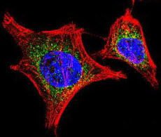
  1. Cells grown in culture:

This was one of the first experiments in my laboratory using fluorescently labelled antibodies, carried out by a student, Emily Hayes, so long ago that she now has a Ph.D., a husband and two children. The cells are endothelial cells (that line blood vessels). Blue: nuclei; green: F-actin; red: Von Willebrand factor, a protein marker for endothelium.


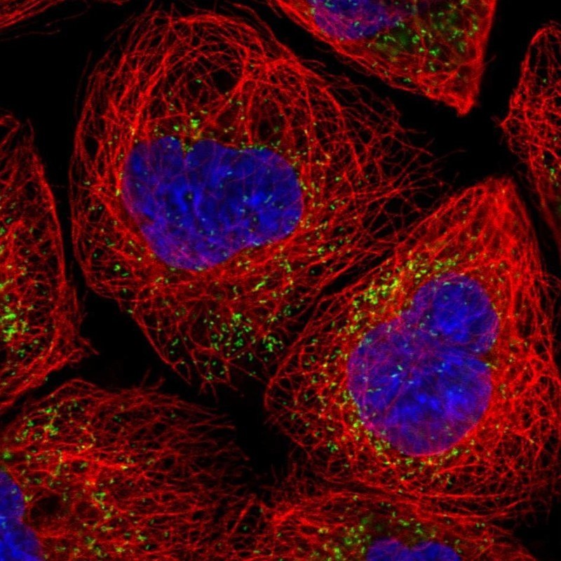
  1. Two very recent images taken by my colleague Roderik Kortlever of a senescent mouse fibroblast and of mouse breast tissue:


3. Waves of calcium in firing neurons:

One of my fondest memories is helping to do the first experiment that measured the level of calcium within a cell, carried out with my colleague the late Roger Tsien and two other friends. I only grew the cells: Roger had designed and made the molecule, quin2. We didn’t know it at the time but Roger’s wonder molecule was the first of many intracellular ‘reporters.’ Roger shared the 2008 Nobel Prize in Chemistry for his discovery and development of the green fluorescent protein with organic chemist Osamu Shimomura and neurobiologist Martin Chalfie.

This wonderful video of a descendant of quin2 in nerve cells was made in Dr. Sakaguchi’s lab at Iowa State University.


4. Calcium wave flooding a fertilized egg: Taro Kaneuchi and colleagues at the Tokyo Metropolitan University:

Click for a time-lapse movie of an egg cell that has been artificially stimulated to show the kind of calcium change that happens at fertilization. In this time-lapse movie the calcium level reaches a maximum signal intensity after about 30 min before gradually decreasing to the basal level.


5. The restless cell (1):

This movie shows how protein filaments in cells can continuously break down and reform – called treadmilling. Visualised in HeLa cells using a green fluorescent protein that sticks to microtubules (tubular polymers made up of the protein tubulin) by HAMAMATSU PHOTONICS.


6. The restless cell (2):

This movie shows how mitochondria (organelles within the cell) are continuously changing shape and moving within the cell’s interior (cytosol). Red marks the mitochondria; green DNA within the nucleus. HAMAMATSU PHOTONICS.


7. Cell division:

Pig kidney cells undergoing mitosis. Red marks DNA (nucleus); green is tubulin: HAMAMATSU PHOTONICS.


8. DNA portrait of Sir John Sulston by Marc Quinn commissioned by the National Portrait Gallery:This image looks a bit drab in the present context but in some ways it’s the most dramatic of all. John Sulston shared the 2002 Nobel Prize in Physiology or Medicine with Sydney Brenner and Robert Horvitz for working out the cell lineage of the roundworm Caenorhabditis elegans (i.e. how it develops from a single, fertilized egg to an adult). He went on to sequence the entire DNA of C. elegans. Published in 1998, it was the first complete genome sequence of an animal — an important proof-of-principle for the Human Genome Project that followed and for which Sulston directed the British contribution at the Sanger Centre in Cambridgeshire, England. The project was completed in 2003.

The portrait shows colonies of bacteria in a jelly that, together, carry all Sulston’s DNA. This represents DNA cloning in which DNA fragments, taken up by bacteria after insertion into a circular piece of DNA (a plasmid), are multiplied to give many identical copies for sequencing.


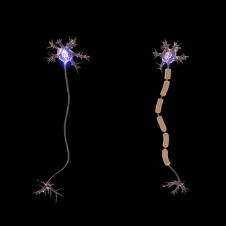
9. “Brainbow” mice by Tamily Weissman at Harvard University:

The science behind this astonishing image builds on the work of Roger Tsien. Mice are genetically engineered to carry three different fluorescent proteins corresponding to the primary colours red, yellow and blue. Within each cell recombination occurs randomly, giving rise to different colours. The principle of mixing primary colours is the same as used in colour televisions.  In this view individual neurons in the brain (specifically a layer of the hippocampus) project their dendrites into the outer layer. Other magnificent pictures can be seen in the Cell Picture Show.

It’s certainly science – but is it art?

A few years ago the Fitzwilliam Museum in Cambridge staged Vermeer’s Women, an exhibition of key works by Johannes Vermeer and over thirty other masterpieces from the Dutch ‘Golden Age’. I tried the experiment of standing in the middle of each room and picking out the one painting that, from a distance, most caught my amateur eye. Funny thing was: not one turned out to be by the eponymous star of the show! Wondrous though Vermeer’s paintings were, the ones that really took my fancy were by Pieter de Hooch, Samuel van Hoogstraten and Nicolaes Maes, guys I’d never heard of.

Which made the point that you don’t need to be a big cheese to make a splash and that in the new Dutch Republic of the 17th century, the most prosperous nation in Europe, there was enough money to keep a small army of splodgers in palettes and paint. Skillful and incredibly patient though these chaps were, they simply used the tools available to paint what they saw in the world before them — as for the most part have artists down the ages.

But hang on! Isn’t that what we’ve just been on about? Scientists applying enormous skill and patience in using the tools they’ve developed to visualize life — to image what Nature lays before them. So the only difference between the considerable army of biological scientists around the world making a new art form and the Old Masters is that the newcomers are unveiling life — as opposed to the immortalizing a rather dopy-looking aristocrat learning to play the virginal or some-such.

Controversial?

Not really. Let’s leave the last word to Roger Tsien. In our final picture there are eight bacterial colonies each expressing a different colour of fluorescent protein arranged to grow as a San Diego beach scene in a Petri dish. It became the logo of Roger’s laboratory.

Lorenzo’s Oil for Nervous Breakdowns


A Happy New Year to all our readers – and indeed to anyone who isn’t a member of that merry band!

What better way to start than with a salute to the miracles of modern science by talking about how the lives of a group of young boys have been saved by one such miracle.

However, as is almost always the way in science, this miraculous moment is merely the latest step in a long journey. In retracing those steps we first meet a wonderful Belgian – so, when ‘name a famous Belgian’ comes up in your next pub quiz, you can triumphantly produce him as a variant on dear old Eddy Merckx (of bicycle fame) and César Franck (albeit born before Belgium was invented). As it happened, our star was born in Thames Ditton (in 1917: his parents were among the one quarter of a million Belgians who fled to Britain at the beginning of the First World War) but he grew up in Antwerp and the start of World War II found him on the point of becoming qualified as a doctor at the Catholic University of Leuven. Nonetheless, he joined the Belgian Army, was captured by the Germans, escaped, helped by his language skills, and completed his medical degree.

Not entirely down to luck

This set him off on a long scientific career in which he worked in major institutes in both Europe and America. He began by studying insulin (he was the first to suggest that insulin lowered blood sugar levels by prompting the liver to take up glucose), which led him to the wider problems of how cells are organized to carry out the myriad tasks of molecular breaking and making that keep us alive.

The notion of the cell as a kind of sac with an outer membrane that protects the inside from the world dates from Robert Hooke’s efforts with a microscope in the 1660s. By the end of the nineteenth century it had become clear that there were cells-within-cells: sub-compartments, also enclosed by membranes, where special events took place. Notably these included the nucleus (containing DNA of course) and mitochondria (sites of cellular respiration where the final stages of nutrient breakdown occurs and the energy released is transformed into adenosine triphosphate (ATP) with the consumption of oxygen).

In the light of that history it might seem a bit surprising that two more sub-compartments (‘organelles’) remained hidden until the 1950s. However, if you’re thinking that such a delay could only be down to boffins taking massive coffee breaks and long vacations, you’ve never tried purifying cell components and getting them to work in test-tubes. It’s a process called ‘cell fractionation’ and, even with today’s methods, it’s a nightmare (sub-text: if you have to do it, give it to a Ph.D. student!).

By this point our famous Belgian had gathered a research group around him and they were trying to dissect how insulin worked in liver cells. To this end they (the Ph.D. students?!) were using cell fractionation and measuring the activity of an enzyme called acid phosphatase. Finding a very low level of activity one Friday afternoon, they stuck the samples in the fridge and went home. A few days later some dedicated soul pulled them out and re-measured the activity discovering, doubtless to their amazement, that it was now much higher!

In science you get odd results all the time – the thing is: can you repeat them? In this case they found the effect to be absolutely reproducible. Leave the samples a few days and you get more activity. Explanation: most of the enzyme they were measuring was contained within a membrane-like barrier that prevented the substrate (the chemical that the enzyme reacts with) getting to the enzyme. Over a few days the enzyme leaked through the barrier and, lo and behold, now when you measured activity there was more of it!

Thus was discovered the ‘lysosome’ – a cell-within-a cell that we now know is home to an array of some 40-odd enzymes that break down a range of biomolecules (proteinsnucleic acidssugars and lipids). Our self-effacing hero said it was down to ‘chance’ but in science, as in other fields of life, you make your own luck – often, as in this case, by spotting something abnormal, nailing it down and then coming up with an explanation.

In the last few years lysosomes have emerged as a major player in cancer because they help cells to escape death pathways. Furthermore, they can take up anti-cancer drugs, thereby reducing potency. For these reasons they are the focus of great interest as a therapeutic target.

Lysosomes in cells revealed by immunofluorescence.

Antibody molecules that stick to specific proteins are tagged with fluorescent labels. In these two cells protein filaments of F-actin that outline cell shape are labelled red. The green dots are lysosomes (picked out by an antibody that sticks to a lysosome protein, RAB9). Nuclei are blue (image: ThermoFisher Scientific).

Play it again Prof!

In something of a re-run of the lysosome story, the research team then found itself struggling with several other enzymes that also seemed to be shielded from the bulk of the cell – but the organelle these lived in wasn’t a lysosome – nor were they in mitochondria or anything else then known. Some 10 years after the lysosome the answer emerged as the ‘peroxisome’ – so called because some of their enzymes produce hydrogen peroxide. They’re also known as ‘microbodies’ – little sacs, present in virtually all cells, containing enzymatic goodies that break down molecules into smaller units. In short, they’re a variation on the lysosome theme and among their targets for catabolism are very long-chain fatty acids (for mitochondriacs the reaction is β-oxidation but by a different pathway to that in mitochondria).

Peroxisomes revealed by immunofluorescence.

As in the lysosome image, F-actin is red. The green spots here are from an antibody that binds to a peroxisome protein (PMP70). Nuclei are blue (image: Novus Biologicals)

Cell biology fans will by now have worked out that our first hero in this saga of heroes is Christian de Duve who shared the 1974 Nobel Prize in Physiology or Medicine with Albert Claude and George Palade.

A wonderful Belgian. Christian de Duve: physician and Nobel laureate.

Hooray!

Fascinating and important stuff – but nonetheless background to our main story which, as they used to say in The Goon Show, really starts here. It’s so exciting that, in 1992, they made a film about it! Who’d have believed it?! A movie about a fatty acid!! Cinema buffs may recall that in Lorenzo’s Oil Susan Sarandon and Nick Nolte played the parents of a little boy who’d been born with a desperate disease called adrenoleukodystrophy (ALD). There are several forms of ALD but in the childhood disease there is progression to a vegetative state and death occurs within 10 years. The severity of ALD arises from the destruction of myelin, the protective sheath that surrounds nerve fibres and is essential for transmission of messages between brain cells and the rest of the body. It occurs in about 1 in 20,000 people.

Electrical impulses (called action potentials) are transmitted along nerve and muscle fibres. Action potentials travel much faster (about 200 times) in myelinated nerve cells (right) than in (left) unmyelinated neurons (because of Saltatory conduction). Neurons (or nerve cells) transmit information using electrical and chemical signals.

The film traces the extraordinary effort and devotion of Lorenzo’s parents in seeking some form of treatment for their little boy and how, eventually, they lighted on a fatty acid found in lots of green plants – particularly in the oils from rapeseed and olives. It’s one of the dreaded omega mono-unsaturated fatty acids (if you’re interested, it can be denoted as 22:1ω9, meaning a chain of 22 carbon atoms with one double bond 9 carbons from the end – so it’s ‘unsaturated’). In a dietary combination with oleic acid  (another unsaturated fatty acid: 18:1ω9) it normalizes the accumulation of very long chain fatty acids in the brain and slows the progression of ALD. It did not reverse the neurological damage that had already been done to Lorenzo’s brain but, even so, he lived to the age of 30, some 22 years longer than predicted when he was diagnosed.

What’s going on?

It’s pretty obvious from the story of Lorenzo’s Oil that ALD is a genetic disease and you will have guessed that we wouldn’t have summarized the wonderful career of Christian de Duve had it not turned out that the fault lies in peroxisomes.

The culprit is a gene (called ABCD1) on the X chromosome (so ALD is an X-linked genetic disease). ABCD1 encodes part of the protein channel that carries very long chain fatty acids into peroxisomes. Mutations in ABCD1 (over 500 have been found) cause defective import of fatty acids, resulting in the accumulation of very long chain fatty acids in various tissues. This can lead to irreversible brain damage. In children the myelin sheath of neurons is damaged, causing neurological defects including impaired vision and speech disorders.

And the miracle?

It’s gene therapy of course and, helpfully, we’ve already seen it in action. Self Help – Part 2 described how novel genes can be inserted into the DNA of cells taken from a blood sample. The genetically modified cells (T lymphocytes) are grown in the laboratory and then infused into the patient – in that example the engineered cells carried an artificial T cell receptor that enabled them to target a leukemia.

In Gosh! Wonderful GOSH we saw how the folk at Great Ormond Street Hospital adapted that approach to treat a leukemia in a little girl.

Now David Williams, Florian Eichler, and colleagues from Harvard and many other centres around the world, including GOSH, have adapted these methods to tackle ALD. Again, from a blood sample they selected one type of cell (stem cells that give rise to all blood cell types) and then used genetic engineering to insert a complete, normal copy of the DNA that encodes ABCD1. These cells were then infused into patients. As in the earlier studies, they used a virus (or rather part of a viral genome) to get the new genetic material into cells. They choose a lentivirus for the job – these are a family of retroviruses (i.e. they have RNA genomes) that includes HIV. Specifically they used a commercial vector called Lenti-D. During the life cycle of RNA viruses their genomes are converted to DNA that becomes a permanent part of the host DNA. What’s more, lentiviruses can infect both non-dividing and actively dividing cells, so they’re ideal for the job.

In the first phase of this ongoing, multi-centre trial a total of 17 boys with ALD received Lenti-D gene therapy. After about 30 months, in results reported in October 2017, 15 of the 17 patients were alive and free of major functional disability, with minimal clinical symptoms. Two of the boys with advanced symptoms had died. The achievement of such high remission rates is a real triumph, albeit in a study that will continue for many years.

In tracing this extraordinary galaxy, one further hero merits special mention for he played a critical role in the story. In 1999 Jesse Gelsinger, a teenager, became the first person to receive viral gene therapy. This was for a metabolic defect and modified adenovirus was used as the gene carrier. Despite this method having been extensively tested in a range of animals (and the fact that most humans, without knowing it, are infected with some form of adenovirus), Gelsinger died after his body mounted a massive immune response to the viral vector that caused multiple organ failure and brain death.

This was, of course, a huge set-back for gene therapy. Despite this, the field has advanced significantly in the new century, both in methods of gene delivery (including over 400 adenovirus-based gene therapy trials) and in understanding how to deal with unexpected immune reactions. Even so, to this day the Jesse Gelsinger disaster weighs heavily with those involved in gene therapy for it reminds us all that the field is still in its infancy and that each new step is a venture into the unknown requiring skill, perseverance and bravery from all involved – scientists, doctors and patients. But what better encouragement could there be than the ALD story of young lives restored.

It’s taken us a while to piece together the main threads of this wonderful tale but it’s emerged as a brilliant example of how science proceeds: in tiny steps, usually with no sense of direction. And yet, despite setbacks, over much time, fragments of knowledge come together to find a place in the grand jigsaw of life.

In setting out to probe the recesses of metabolism, Christian de Duve cannot have had any inkling that he would build a foundation on which twenty-first century technology could devise a means of saving youngsters from a truly terrible fate but, my goodness, what a legacy!!!

References

Eichler, F. et al. (2017). Hematopoietic Stem-Cell Gene Therapy for Cerebral Adrenoleukodystrophy. The New England Journal of Medicine 377, 1630-1638.