Two Heads Better Than One

These days the notion that cancer cells have picked up genetic alterations (i.e. in their DNA) and that these change the expression of lots of genes is fairly familiar. But what does ‘gene expression’ really mean and how is it controlled? Well, gene expression is the generation of an RNA molecule that carries the same coding information that is enshrined in the sequence of bases in the DNA of that gene — the process of making the RNA is called transcription. The code carried by most RNAs (messenger RNAs — mRNAs) can subsequently be ‘translated’ into the corresponding sequence of amino acids in a protein. And proteins, of course, do all the work to make cells function.

Self-control

And control? There are two levels: 1: the transcription step is regulated to limit the amount of mRNA made from a particular gene. 2: the translation of mRNA into proteins is regulated by post-transcriptional events.

It’s Step 1 that concerns us today in which proteins bind to specific regulatory sequences of DNA in a gene to modulate the activity of the enzyme that makes mRNA molecules from the DNA template. These proteins are generally called transcription factors.

So to be clear: transcription factors are sequence-specific DNA-binding proteins that control the rate of transcription of genetic information from DNA to mRNA — carried out by the enzyme RNA polymerase II. They regulate — turn on or off — genes so that they are expressed in the right cells at the right time. Groups of transcription factors act in a coordinated way to control all cellular processes (growth, division, etc.) and humans have about 1500 of them.

Regulation of gene expression. This scheme shows the concept of regulatory regions within DNA (enhancers and promoters) coming together to control whether a gene is ‘on’ or ‘off’ as a result of transcription factors and mediator proteins binding to specific sequences. When a  gene is turned on RNA is synthesised by the action of RNA polymerase II. A TATA box is a sequence of DNA found in the core promoter region of genes. Other proteins (coactivatorschromatin remodelershistone acetyltransferaseshistone deacetylaseskinases and methylases) also regulate gene expression but as these lack DNA-binding domains they are not transcription factors.

Transcription factor of the day

It’s handy to be clear about these basics before we turn to an exciting paper by Sai Gourisankar, Gerald Crabtree and colleagues from Stanford University and the MD Anderson Cancer Center, Houston entitled “Rewiring Cancer Drivers”. They focussed on a transcription factor called B-cell lymphoma-6 (BCL6) that plays an important role in the formation of lymphoid tissues and the production of antibodies. BCL6 normally acts as a repressor — it turns genes off. However, as you’ll guess from its name, BCL6 commonly undergoes mutations in various lymphomas, specifically forms of B-cell non-Hodgkin lymphoma.
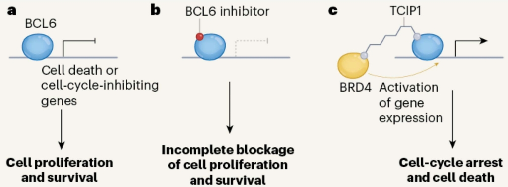
Engineering a transcription factor. a. BCL6 is a transcription factor that, in B cells, normally turns off genes encoding cell cycle inhibitors, thereby allowing some cancer cells to proliferate.  b. Small molecule inhibitors of BCL6 have shown some activity as anti-cancer agents.  c. Chemical coupling of TCIP1to BCL6 recruits BRD4, a transcription activator. This gives strong activation of genes normally turned off by BCL6 and kills cancer cells expressing BCL6. From Phelan and Staudt, 2023.

Why it’s exciting

Scheme c above shows Gourisankar & Co’s clever idea. They coupled BCL6 to a small molecule called TCIP1 (if you’re really interested this stands for transcriptional/epigenetic chemical inducer of proximity). TCIP1 binds to the protein BRD4 (bromodomain-containing protein 4) — a transcriptional and epigenetic regulator that plays a critical role in embryogenesis and cancer development. This juxtaposes BRD4 and BCL6, the upshot being that BRD4 potently activates the expression of genes normally silenced by BCL6.

Great chemistry — so what?

TCIP1 is an extremely potent killer of diffuse large B cell lymphoma cells that express BCL6. Thus the approach of using TCIP1 offers a new therapeutic strategy for these cancers. However, it may also be effective against other cancer types and there is good reason for optimism because there was a bonus in this paper in that TCIP1 also downregulates MYC — a master regulator that is abnormally expressed in many cancers. It’s not clear how this works as BRD4 is generally considered to drive cancers by activating MYC — so the mechanism is indirect but nontheless welcome for that!

And the down-side?

The flip side is that BCL6 suppresses inflammation in immune cells (knocking out BCL6 kills mice via an inflammatory reaction) which might be a problem for a treatment that targets this transcription factor. However, the authors looked hard in both normal mice and in human cells (fibroblasts) for evidence of tissue damage and found none, even though there were large changes in gene expression in spleen and lymphocyte cells.

Clearly there is much to learn about this approach of using what is in effect a double-headed molecule as an anti-cancer agent. The name of the Roman god of beginnings and endings has already been nicked by molecular biologists for the JAK kinases (their two faces link cytokine receptors to other proteins). Let us therefore avoid giving a trendy name to this new approach using TCIP1 but perhaps just nod to Janus in the hope that it will become a useful anti-cancer strategy.

References

Phelan JD, Staudt LM. Double-headed molecule activates cell-death pathways in cancer cells. Nature. 2023 Aug;620(7973):285-286. doi: 10.1038/d41586-023-02213-4. PMID: 37495782.

Gourisankar S, Krokhotin A, Ji W, Liu X, Chang CY, Kim SH, Li Z, Wenderski W, Simanauskaite JM, Yang H, Vogel H, Zhang T, Green MR, Gray NS, Crabtree GR. Rewiring cancer drivers to activate apoptosis. Nature. 2023 Aug;620(7973):417-425. doi: 10.1038/s41586-023-06348-2. Epub 2023 Jul 26. Erratum in: Nature. 2023 Sep;621(7977):E27. PMID: 37495688.

The Jekyll & Hyde of Cancer

If you stood on a street corner and asked passers-by to name a ‘cancer gene’ I suspect p53 would be top choice — setting aside that most people would just gaze at you blankly. That’s because p53 is the most commonly mutated gene in human cancer (as we recently noted in “Be amazed”) — rates varying from 10% to 100%, depending on the cancer type. Approaching 50% of ovarian, oesophageal, bowel and lung cancers lose p53 activity, as do 20 to 40% of breast cancers where it’s loss is associated with more aggressive disease and worse overall survival. So a lot of folk will have heard of it. Oh, and be prepared to buy the drinks if someone says “There’s no such thing as a ‘cancer gene’ — that’s jargon used to mean a gene that is required for normal cell growth and multiplication that has become mutated so that it no longer exerts normal control”.

As it happens, if you posed the same question to cancer biologists you’d probably get the same answer — though we would hope with fewer blank looks and more smart put-downs — because the Web of Science database reveals that over the last 40 years there have been 127,677 separate publications with something to say about this gene.

What do we know?

So what have we gleaned from all that sweat and toil? The gene is TP53 (for tumour protein 53 — the mass of the protein molecule is 53 kilodaltons). It encodes a transcription factor (i.e. a protein that sticks to DNA to control expression of genes). In addition to the full-length protein, the human TP53 gene encodes at least 15 distinct proteins of widely differing size.

TP53 is considered to be a ‘tumour suppressor’ — loss of function of both copies of the gene (and hence the protein) equates to loss of growth control — a major brake on cell proliferation is released. Its normal role is to get turned on when DNA is damaged — p53 expression is exquisitely sensitive to this: just one double-strand break in DNA causes significant amounts of protein to be made. So if p53 can’t be made cells have a green light to reproduce with damaged DNA — a recipe for cancer. But p53 is something of a maverick because mutated versions of one copy of TP53 can produce proteins that interact with the normal (wild-type) protein made from the other gene copy — called a dominant-negative effect. In this way mutant p53 can act to help tumour development. When it comes to mutations p53 is in a class of its own: no fewer than 30,000 have been found and no less than two databases are devoted to listing them. Of these mutations, six amino acids are the most frequent — they’re ‘hotspots’ — and mostly those mutations stop p53 from doing its normal job of ensuring that cells only replicate when they’re in a fit state to do so.

But despite all that molecular stuff, we don’t really know how p53 works and, in a remarkable series of experiments, Eliran Kadosh, Moshe Oren and colleagues from Israel and New York have just revealed another amazing facet of this mysterious gene. They showed that two of the most common mutant p53s can act as stronger tumour suppressors than the normal, wild-type version. They showed this in organoids (3D tissue samples) derived from mouse tissue. However, when they looked in the normal gut in the presence of the usual bacteria (microbiota) these suppressive effects are completely abolished and mutant p53 becomes a tumour driver (i.e. an oncoprotein). Extraordinary!

How did they do it?

They used two mouse models of intestinal cancer — that’s the form of cancer that develops in the small intestines, the plumbing that connects the stomach to the large intestine (the colon). Each model had a mutation in a signalling pathway that can drive malignant transformation, i.e. cancer development. [The genes involved were a kinase (CKIa) that was knocked out and a tumour suppressor (Apc) that was mutated: both manipulations turn on an important signalling pathway (the WNT pathway) and they model human bowel cancer].

The first experiment with CKIa deleted in the gut compared mice expressing normal (wild-type) p53 with one of the ‘hot-spot’ mutations. As expected, knocking out p53 caused pre-cancerous changes and raised cell proliferation throughout the bowel. However, in mice making mutated p53 widely differing effects were seen in different gut segments: the colon and ileum were highly abnormal and proliferative whilst the duodenum and the jejunum were normal.

A possible explanation is that somehow the DNA binding activity of mutant p53 had been restored so that it could now turn on the genes activated by normal p53 that block cell proliferation — but Kadosh & Co showed this had not happened. Mutant p53 remained transcriptionally inert.

As a next step they looked at Apc mutant mice that develop discrete intestinal tumours — these model the human condition familial adenomatous polyposis and tumours typically form in the jejunum with few in the colon. The results followed the CKIa pattern: the expression of mutant p53 increased tumour formation in the colon but reduced it in the jejunum.

Map of human intestines.

Mutant p53 counteracts abnormal growth and tumour development. Photos show different segments of mouse bowel after 5 months with mutated Apc. The effect of mutant p53 (p53R172H i.e. amino acid 172 is changed from R(arginine) to H (histidine)) is to suppress tumour formation in the small intestine (jejunum) but to increase it in the large intestine (colon). Arrows mark polyps — tumour foci. From Kadosh et al. 2020.

The story so far

In mouse models of bowel cancer mutant p53 has contrary effects, blocking tumour growth in the first part of the gut but increasing it in the colon. A stunning result.

What’s going on?

The Kadosh group then carried out a variety of tricky experiments the upshot of which was to show three things:

1. Mutant p53 can suppress genes activated by proteins in the pathway turned on in the mouse models (WNT).

2. Mutant p53 has an even greater tumour-suppressive power than normal (wild-type) p53.

3. Mutant p53 can exert anti-tumour effects not only in the jejunum but also in the ileum, if that is removed from its normal environment.

In casting around for things that might control the activity of mutant p53 the authors alighted on the gut microbiome that has featured frequently in these pages, most recently in “Mushrooming Secret Army”). This comprises an abundance of microorganisms, their number increasing progressively along the gastrointestinal tract (i.e. the jejunum has about 1000 times the duodenum and the colon about one million times that). As we’ve noted in these pages, cross-talk between the microbiome and host cells goes on all the time and upsetting it (e.g., by changing the composition or amount of bugs) is linked to obesity and cancer. Kadosh and colleagues reasoned that microbiota could be responsible for the contrasting effects of mutant versions of p53 in different segments of the gut. They were no doubt delighted when a course of antibiotics released the tumour-suppressive effects of mutant p53 in the colon [i.e. made the colon behave like the jejunum].

They went on to show that mutant 53 did not change the composition of the microbiota in the transgenic mice — strongly suggesting that something released by the bacteria might regulate tumour suppression by mutant p53.

From this it emerged that gallic acid (made by Lactobacillus plantarum and Bacillus subtlilis) could impair the activity of mutant p53. The jejunum (low levels of bacteria) and the ileum (higher levels) have correspondingly different levels of gallic acid. However, treatment of mice with gallic acid caused the duodenum and the jejunum to become highly proliferative and develop many tumour foci (see below). Taken together the results showed that gallic acid, produced by bacteria in both mice and humans but only highly abundant in the colon, completely reproduces the effect of the gut microbiome in abolishing the tumour-suppressive activity of mutant p53.

Gallic acid induces abnormal growth and tumorigenesis when p53 is mutated. Arrows mark polyps. These results in Apc mutant mice were similar to those in CKIa knock-out mice. From Kadosh et al. 2020.

These are extraordinary findings that show not only the power of the microbiome but the amazing flexibility of p53 in that two of its mutant forms, previously considered to have lost tumour suppressive power, can in fact behave as even more powerful suppressors than normal p53. What’s more, their activity is switched from suppressive to oncogenic by a metabolite released from gut bacteria.

Little can Robert Louis Stevenson have guessed that when, back in 1886, he penned his portrait of Dr. Jekyll concocting a potion to suppress his evil urges, he was writing an allegory of the goings on in his own intestines.

Reference

Kadosh, E. et al., (2020). The gut microbiome switches mutant p53 from tumour-suppressive to oncogenic. Nature  586, pages133–138. https://doi.org/10.1038/s41586-020-2541-0.