A Bit of Christmas Cheer

These days we need all the good news going, certainly in the wider world but it also helps in the cancer field. In talking to the general public I’m occasionally brought up short by the question “Why is progress so slow in cancer treatment?” “Well …” I tend to woffle, “It is jolly complicated.” Feeling that, as a scientist, I need to be a bit more precise, I then usually turn to numbers — the statistics of survival, really the bottom line. The statistical folk generally use 5-year survival rates as the timeframe to measure survival rates and for all cancers since 1970 this figure has risen from just under 50% to about 70% — and those figures are pretty much the same for England and the USA.

That’s a significant advance but those figures cloak huge differences between cancer types. Thus major triumphs have seen leukemia survival more than quadruple in the last 50 years, prostate cancer survival tripling and almost all men now surviving testicular cancer (98.2%). However, in the interests of balance we should note that for some cancers (e.g., lung and pancreas) survival rates have shown little improvement — a fact that highlights the immense complexity of cancers.

However, for Christmas let’s return to the bright side and thank Carolyn Taylor and her colleagues at the University of Oxford for a study that followed over half a million women in England who were diagnosed with early invasive breast cancer between January 1993 and December 2015. Their arresting finding is that the number of women who die after a breast cancer diagnosis has decreased by two-thirds since the 1990s.

Fall in the likelihood of dying from breast cancer since the 1990s. From Taylor et al. 2023.

The easiest way to follow this trend is to track vertically from the 5 years since diagnosis point and red off 5, 8, 11 and 15 as the percentage dying in each time block (2010-15, 2005-09, 2000-04 and 1993-99). {‘Cumulative risk’ just means including the combination of all risk factors}.

Think about this in the context of a diagnosis of breast cancer occurring every 10 minutes in the UK. The decrease of two thirds applies to about 55,000 women a year.

That’s a phenomenal improvement and it should brighten Christmas for all of us.

References

Taylor, C. et al. BMJ 2023;381:e074684

USA trends: https://progressreport.cancer.gov/after/survival

Precision CAR-T steering

As you would expect in these pages we have kept up to date with one of the newest, most promising treatments for blood cancer, namely chimeric antigen receptor (CAR)-T cell therapy (Cardiff Crock of Gold? And Gosh! Wonderful GOSH).

What is CAR-T cell therapy?

As a reminder, the idea is that cells are taken from a patient’s blood and a specific population (T cells, part of the immune system) is genetically engineered in the laboratory to improve their efficiency in attacking cancer cells. It’s been used to treat blood cancers (lymphomas, leukemias and multiple myeloma). In several large trials it’s been shown to be more effective than the standard treatment for patients with non-Hodgkin lymphoma and for chronic lymphocytic leukaemia. The response rates have slowly risen and now about half of treated patients respond to CAR-T cell therapy.

In Gosh! Wonderful GOSH we described how ‘designer immune cells’ were made to improve the efficiency with which the T cells target a particular protein (CD19), present on the surface of leukemia cells, by giving them artificial T cell receptors (these are ‘chimeric T cell receptors’ or ‘chimeric antigen receptors (CARs)’ – because they’re made by fusing several bits together to make something that sticks to the target ‘antigen’ – CD19). The engineered receptors thereby boost the immune response against the leukemia.

You Lu at the West China Hospital in Chengdu went a step further by taking immune cells from people with aggressive lung cancer and disabling the PDCD1 (usually called PD-1) gene (Cardiff Crock of Gold?) The PD-1 protein normally attenuates the immune system to prevent it attacking its own tissues but, as this reduces its anti-cancer capacity, knocking out PD-1 should overcome that restriction.

Sharpening CRISPR

Things have just taken another step forward thanks to Jiqin Zhang and colleagues at institutes in Shanghai and Hangzhou, China who found a way to make targeted CAR-T cells without using viruses. The concern here is that genetic material carried by viruses can integrate randomly into the genome (DNA) of T cells and, in the worst scenario, this may actually promote cancer. What Zhang & Co did was to use the DNA-editing tool CRISPR–Cas9 to engineer precisely CAR-T cells without using viruses. Happily we have explained CRISPR in previous pieces and in Sharpening CRISPR we noted how genome editing can be achieved using parts of CRISPR together with other enzymes to insert point mutations directly without making breaks in double-stranded in DNA.

Specifically, they used this method to make CAR-T cells carrying the CD19-specific antigen — as done previously. However, the really cunning bit was that they inserted the CD19-binding sequence into the PD-1 gene. This produced what they called PD1-19bbz cells that bear the CD19 antigen but do not express the T-cell-suppressing protein PD-1.

These cells, when injected intravenously in to mice, produced spectacular effects on tumours (see figure below). To visualise the human-derived tumours the cells were engineered to be bioluminescent (a method for live tissue imaging in animals). Within 7 days of CAR-T cell infusion the blue luminescence indicating tumour cells has completely disappeared.

Human lymphoma cell growth blocked by PD1-19bbz cells. Shown are images of 8 untreated mice (left) at Days 1 and 7 and of 8 mice (right) 14 days after CAR-T cell infusion at Day1. The purple colour is the bioluminescence signal from the tumour cells showing that by Day 7 untreated mice are completely overwhelmed by the tumour whereas treated mice remain tumour free for 28 days (shown at 14 days). From Zhang et al., 2022.

And in humans?

PD1-19bbz cells gave complete remission in seven people in response to the virus-free, precisely engineered CAR-T cells without serious adverse events.

This two-in-one approach is a significant advance because not only is it safer but the production of cells modified in this way is simpler than by viral methods and offers an innovative technology for CAR-T cell therapy.

‘Personalized medicine’ … at last!!

Hard on the heels of Zhang et al comes an update from Susan Foy, Antoni Ribas and colleagues at PACT Pharma, South San Francisco and various branches of the University of California, who have carried out a small clinical trial (16 people with solid tumours including breast and colon) — of what’s been described as “the most complicated therapy ever”. Rather than trying to boost the immune response via CD19 signalling, they targeted the T cell receptor (TCR) that confers on T cells the exquisite specificity of mutation recognition. The idea was to use CRISPR/Cas9 non-viral precision genome editing (just as Jiqin Zhang did) to knock-out simultaneously the two endogenous TCR genes, TCRα (TRAC) and TCRβ (TRBC), and insert in the TRAC locus the two chains of a neoantigen-specific TCR (neoTCR), isolated from the patient’s own circulating T cells using a personalized library of soluble predicted neoantigen-HLA capture reagents (recall that neoantigens and their antigenic determinants — the neoepitopes —  are processed and presented by human leukocyte antigen (HLA) to be recognized by T cells and generate antibodies. Algorithms predicted which mutations were most likely to provoke a T cell response.

After one month of treatment in five of the 16 participants the cancer had stabilised (the tumours had not grown), with only two experiencing side effects probably caused by the activity of the edited T cells. That may sound like a low ‘success’ rate but this trial was really a ‘proof of principle’. It showed that the method is safe to use in patients and, as one of the authors said, “We just need to hit it stronger the next time.”

The authors noted that it was a formidably time-consuming effort — they had to start by sequencing DNA from blood samples and tumour biopsies, then selecting the neo-antigen mutations (in the tumour but not blood) — for each patient.

It is, however, a tremendous step forward and, yes, there’s plenty of room for improvements

References

Zhang, J., Hu, Y., Yang, J. et al. Non-viral, specifically targeted CAR-T cells achieve high safety and efficacy in B-NHL. Nature (2022). https://doi.org/10.1038/s41586-022-05140-y.

Foy, S.P., Jacoby, K., Bota, D.A. et al. Non-viral precision T cell receptor replacement for personalized cell therapy. Nature (2022). https://doi.org/10.1038/s41586-022-05531-1

Cockles and Mussels, Alive, Alive-O!

And so they are across the globe, not forgetting clams, a term that can cover all bivalve molluscs – a huge number of species (over 15,000), all having a two-part, hinged shell. The body inside doesn’t have a backbone, making it soft and edible on a scale of keeping-you-alive to orgasmic, depending on the consumer – oysters and scallops are part of the family.

Bivalves are particularly common on rocky and sandy coasts where they potter happily along, generally burrowing into sediment although some of them, scallops for instance, can swim. By and large their only problem is that humans like to eat them.

Clamming up

However, it gradually emerged in the 1970s that there was another cloud hovering over some of these gastronomic delights. Their commercial importance had drawn attention to the fact that soft-shell clams living along the east coast of North America, together with mussels on the west coast and cockles in Ireland, were dying in large numbers. The cause was an unusual type of cancer in which leukemia-like cells reproduce until they turn the blood milky and the animals die, in effect, from asphyxiation. In soft-shell clams, also known as sand gapers and steamers, the disease has spread over 1,500 km from Chesapeake Bay to Prince Edward Island.

A 2009 study had shown that as the disease progresses there is a rise in the number of blood cells that have abnormally high amounts of DNA (in clams typically four times the normal number of chromosomes – i.e. they’re tetraploid). In parallel with this change the cells make increasing amounts of an enzyme called reverse transcriptase (RT).

That was pretty surprising as RT does what its name suggests: reverses part of the central dogma of molecular biology (DNA makes RNA makes protein) by using RNA as a template to make DNA. RT is usually carried by viruses whose hereditary material is RNA (rather than DNA – so they’re called retroviruses). As part of their life cycle they turn their genomes into DNA that inserts into the host’s genome – which gets reproduced (as RNA) to make more viruses.

But how did RT get into clams? Enter Michael Metzger and Stephen Goff from Columbia University in New York, together with Carol Reinisch and James Sherry from Environment Canada, who began to unravel the mystery.

Jumping genes

Using high throughput sequencing they showed that clam genomes contain stretches of about 5,000 bases that came about when the RNA of a virus was copied into DNA by RT (reverse transcriptase) and then inserted into the host chromosome. Normal clams have from two to ten copies of this ‘repetitive element’ that Metzger & Co dubbed Steamer. That wasn’t too surprising as we have repetitive DNA too – it makes up about half the human genome. Many of these repeated sequences can move around within the genome – they’re often called ‘jumping genes’ – and it’s easy to see how this can happen when RT uses RNA to make DNA that can then pop into new sites in the genome. And you might guess that this process could damage the host DNA in ways that might lead to disease.

A long jump?

It turned out that the diseased clams had suffered massive amplification of Steamer to the extent that they carry 150 to 300 copies of the sequence. So that’s about 30 times as many Steamer DNAs being scattered across the clam genome – but how could that cause the same disease all the way from New York to Prince Edward Island? The answer came from peering into the DNA sequences of the tumour cells: they were virtually identical to each other – but they were different to those of their hosts! Meaning? The damage that led to leukemia, caused by shoe-horning 100s of extra copies of Steamer into clam genomes, only occurred once. And the staggering implication of that finding is that the cancer spread from a single ‘founder’ clam throughout these marine-dwelling molluscs. The resemblance to the way the cancer spreads in Tasmanian devils is striking.

Fishier and fishier

Fast forward to June 2016 and the latest contribution from the Metzger group reporting four more examples of transmissible cancer in bivalves – in mussels from British Columbia, in golden carpet shell clams from the Spanish coast and two forms in cockles.

Each appears to cause the same type of leukemia previously found in clams. The disease appears to be transmitted ‘horizontally’, i.e. by living cancer cells, descended from a single common ancestor, passing directly from one animal to another. Indeed, if you transplant blood cells from infected animals into normal clams they get leukemia.

 Species hopping

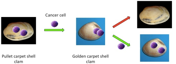
All that is quite amazing but the genetic analysis came up with an even more bizarre finding. In the golden carpet shell clams DNA from cancer cells showed no match with normal DNA from this species. It was clearly derived from a different species, which turned out to be the pullet shell clam – a species that, by and large doesn’t get cancer. So they have presumably come up with a way of resisting a cancer that arose in them, whilst at the same time being able to pass live tumour cells on to another species!!clam-transfer-pic

Cancer cell transmission between different species of shellfish. Cancer cells can arise in one species (pullet shell clams) that do not themselves develop leukemia but are able to pass live cells to another species (golden carpet shell clams) that do get leukemia (Metzger et al. 2016).

We have no idea how the cancer cells survive transfer. It seems most likely that they are taken up through the siphons that molluscs use for feeding, respiration, etc. and then somehow get across the walls of the respiratory/digestive systems. In the first step they would have to survive exposure to sea water which contains a lot more salt than cells are happy in. The ‘isotonic’ saline used in drips to infuse patients contains 0.9% salt whereas seawater, with 3.5%, is ‘hypertonic’ – cells put in a hypertonic solution will shrink as water is drawn out of the cell into the surrounding solution. Presumably the cells shrivel up a bit but some at least take this in their stride and recover to reproduce in their new host. Equally obscure is how a species can protect itself from a cancer that it can pass to another species.

These amazing findings throw a different light on the care-free underwater life depicted in Disney’s The Little Mermaid, in which the popular song ‘Under the Sea’ fails to mention floating cancer.

Can this happen to us?!!

Well, not as far as we know. But the fact that the known number of cancers that can be passed from one animal to another has now risen to nine does make you wonder. However, there’s no evidence that it happens in humans in anything like the normal course of events. There are examples of person-to-person transfer, notably during organ transplantation, and there is one recent case of cancerous cells from a tapeworm colonising a human host. But these are very rare, the latter occurring in a patient with a severely weakened immune system, and there is no example of spread beyond two people.

Phew! What a relief! So now we can concentrate on following developments both in Tasmania and beneath the waves in the hope that, not only can we go on satisfying our lust for clam bakes and chowders, but that these incredible creatures will reveal secrets that will benefit mankind.

References

AboElkhair, M. et al. (2009). Reverse transcriptase activity associated with haemic neoplasia in the soft-shell clam Mya arenaria. Diseases of Aquatic Organisms 84, 57-63.

Arriagada, G. et al. (2014). Activation of transcription and retrotransposition of a novel retroelement, Steamer, in neoplastic hemocytes of the mollusk Mya arenaria. PNAS 2014 111 (39) 14175-14180; published ahead of print September 8, 2014, doi:10.1073/pnas.1409945111.

Metzger, M.J. et al. (2015). Horizontal Transmission of Clonal Cancer Cells Causes Leukemia in Soft-Shell Clams. Cell 161, 255–263.

Metzger, M.J. et al. (2016). Widespread transmission of independent cancer lineages within multiple bivalve species. Nature 534, 705–709.

Muehlenbachs, A. et al. (2015). Malignant Transformation of Hymenolepis nana in a Human Host. N Engl J Med 2015; 373:1845-1852.

Keeping Cancer Catatonic

Over a century ago there lived in London an astute physician by the name of Stephen Paget. He was one of those who may or may not be envied in being part of a super-talented family. His Dad, Sir James Paget, was pals with Charles Darwin and, together with Rudolph Virchow, laid the foundations of modern pathology, though today medical students usually encounter his infinitesimal immortality through several diseases that bear his name. These include a rare condition, Paget’s disease of the breast, in which malignant cells form in the skin of the nipple creating an itchy rash, usually treatable by surgery. His Uncle George had been Regius Professor of Physic at Cambridge and he had several brothers, two of whom became bishops. Fortunately Stephen continued the medical thread of the family and Paget’s passion became breast cancer.

A Key Question

Paget had that invaluable scientific gift of being able to pinpoint a key question – in his case ‘What is it that allows tumour cells to spread around the body?’ – and it was such a good question that to this day we don’t have a complete answer. That it happens had been known long before the appearance of Paget Junior. René-Théophile-Hyacinthe Laënnec, French of course, in the early years of the 19th century described how skin cancer could spread to the lungs before he went on to invent the stethoscope in 1816. The mother of this invention was a young lady whom he described as having a ‘great degree of fatness’ that made her heartbeat inaudible by the then conventional method of placing ear to chest. Using a piece of paper rolled into a tube as a bridge, Laënnec was somewhat taken aback that the beat was more distinct than he’d ever heard before. Needless to say, medicine being a somewhat reactionary profession, not all its practitioners had ears tuned to receive this advance with glee but in the end, of course, it caught on and we can therefore award Laënnec first prize in reducing human cumulative embarrassment. It was another French surgeon, Joseph Récamier, who subsequently coined the term metastasis, (to be precise ‘métastase’) to describe the formation of secondary growths derived from a primary tumour.

Early Ideas about Metastasis

The notion that primary tumours could give rise to a diaspora gradually took root but it was not until 1840 that the Munich-born surgeon Karl Thiersch showed that it was actually cells – malignant cells – that wandered off and found new homes. Rudolf Virchow had come up with the idea that spreading was via a ‘juice’ released by primaries that somehow converted normal cells at other sites into tumours. As Virchow was jolly famous, having not only made the study of disease into a science but also discovered leukemia, it took a while for Thiersch to triumph, notwithstanding the evidence of Laënnec and others. Funnily enough, and as quite often happens in scientific arguments, it now looks as though both were right if for ‘juice’ you substitute ‘messengers’ – that is, chemicals dispatched by tumour cells – as we shall see.

Paget’s attention had been drawn to this subject through his observations on breast cancer, and he’d taking as a starting point the most obvious question: ‘How do tumour cells know where to stick?’ Or, as he elegantly phrased it in a landmark paper of 1889: ‘What is it that decides what organs shall suffer in a case of disseminated cancer?’ The simplest answer would be that it just depends on anatomy: when cells leave a tumour and get into the circulation they stick to the first tissue they meet. But in looking at over 700 cases he’d found this just didn’t happen and that secondary growths often appeared in the lungs, kidneys, spleen and bone. Paget acknowledged the uncommonly prescient suggestion a few years earlier by Ernst Fuchs that certain organs may be ‘predisposed’ for secondary cancer and concluded that ‘the distribution of secondary growths was not a matter of chance.’ This led him to a botanical analogy for tumour metastasis: ‘When a plant goes to seed, its seeds are carried in all directions; but they can only live and grow if they fall on congenial soil.’ From this, then, emerged the ‘seed and soil’ theory of metastasis, its great strength being the image of interplay between tumour cells and normal cells, their actions collectively determining the outcome. Rather charmingly, Paget concluded his paper with: ‘The best work in the pathology of cancer is now done by those who are studying the nature of the seed. They are like scientific botanists; and he who turns over the records of cases of cancer is only a ploughman, but his observation of the properties of the soil may also be useful.’

BOOKMARKING

Bookmarking cancer: Primary tumours mark sites around the body to which they will spread (metastasize) by sending out chemical signals that create sticky ‘landing sites’ (red protein A) on target cells. Cells released from the bone marrow carry proteins B and C. B attaches to A and tumour cells ‘land’ on C. Cells may remain quiescent in a new site for years or decades, their growth suppressed by signals (e.g., TSP-1) released from nearby blood vessels. Only when appropriate activating signals dominate (e.g., TGFbeta) is secondary tumour growth switched on.

Finding a Landing Strip

For well over a century Paget’s aphorism of  ‘seed and soil’ pretty well summed up our knowledge of metastasis. It’s obvious that before any rational therapy can be designed we need to unravel the molecular detail but we’ve had to wait until the twenty-first century for any further significant insight into the process. As so often in science, the hold-up has been largely due to waiting for the appropriate combination of methods to be developed – in this case fluorescently tagged antibodies to detect specific proteins in cells and tissues and genetically modified mice.

In the forefront of this pursuit has been David Lyden and his colleagues at Weill Cornell Medical College and other centers and their most extraordinary finding is that cells in the primary tumour release proteins into the circulation and these, in effect, tag what will become landing points for wandering cells. Extraordinary because it means that these sites are determined before any tumour cells actually set foot outside the confines of the primary tumour. These are chemical messengers rather equivalent to Virchow’s ‘juice’: they don’t change normal cells into tumour cells but they do direct operations. However, it’s a bit more complicated because, in addition to sending out a target marker, tumours also release proteins that signal to the bone marrow. This is the place where the cells that circulate in our bodies (red cells, white cells, etc.) are made from stem cells. The arrival of signals from the tumour causes some cells to be released into the circulation; these carry two protein markers on their surface: one sticks to the pre-marked landing site, the other to tumour cells once they appear in the circulation. It’s a double-tagging process: the first messenger makes a sticky patch for bone marrow cells that appear courtesy of another messenger, and they become the tumour cell target. It’s molecular Velcro: David Lyden calls it ‘cellular bookmarking.’

Controlling Metastatic Takeoff

Tumour cells that find a new home in this way, after they’ve burrowed out of the circulation, could in principle then take off, growing and expanding as a ‘secondary.’ However, and perhaps surprisingly, generally they do the exact opposite: they go into a state of hibernation, remaining dormant for months or years until some trigger finally sets them off. The same group has now modeled this ‘pre-metastatic niche’ for human breast cancer cells, showing that the switch between dormancy and take-off is controlled by proteins released by nearby blood vessels. The critical protein that locks tumour cells into hibernation appears to be TSP-1 (thrombospondin-1). As long as TSP-1 is made by the blood vessel cells metastatic growth is suppressed. This effect is overridden by stimuli that turn on new vessel growth and in so doing switch secretion from TSP-1 to TGFB (transforming growth factor beta). Now proliferation of the disseminated tumour cells is activated and the micro-metastasis becomes fully malignant. It should be said that this is a model system and may possibly bear little relation to what goes on in real tumours. However, the fact that specific proteins that are, moreover, highly plausible candidates, can control such a switch strongly suggests its relevance and also highlights potential targets for therapeutic manipulation.

Stranger Than Fiction

The system for directing tumour cells to a target seems extraordinarily elaborate. Given that tumour cells cannot evolve in the sense of getting better at being metastatic – they just have to go with what they’ve got – how on earth might it have come about? We don’t know, but the most likely explanation is that they are taking advantage of natural defense mechanisms. Although tumours start from normal cells, the first reaction of the body is to see them as ‘foreign’ – much as it does bugs that get into a cut – and the response is to switch on inflammation and an immune response to eliminate the ‘invader.’

Perhaps what is happening in these mouse models is that the proteins released by the tumour cells are just a by-product of the genetic disruption in cancer cells. Nevertheless, they may signal ‘damage somewhere in the body’. That at least would explain why the bone marrow decides to release cells that are, in effect, a response to the tumour. The second question is trickier: Why should tumours release proteins that mark specific sites? We’ve known since Dr. P’s studies that cells from different tumours do indeed head for different places and it may just be that the messengers arising in the genetic mayhem happen to reflect the tissue of origin. The mouse models, encouragingly, show that the target changes with tumour type (e.g., swap from breast to skin and the cells go somewhere else). In other words, tumours send out their own protein messengers that set up sticky landing strips in different places around the circulation.

As for take-off, it may be that newly arrived tumour cells simply adapt to the style of their neighborhood. By and large, the blood vessels are pretty static structures: they don’t go in for cell proliferation unless told to do so by specific signals, as happens when you get injured and need to repair the damage. TSP-1 appears to be a ‘quiescence’ signal, telling cells to sit tight. The switch to proliferation comes when that signal is overcome by TGFB, activating both blood vessels and tumour. All of which would delight Paget: not only is our expanding picture consistent with ‘seed and soil’ but the control by local signals over what happens next makes his rider that ‘observation of the properties of the soil may also be useful’ spot on.

References

Kaplan, R.N., Riba, R.D., Zacharoulis, S., Bramley, A.H., Vincent, L., Costa, C., MacDonald, D.D., Jin, D.K., Shido, K., Kerns, S.A., Zhu, Z., Hicklin, D., Wu, Y., Port, J.L., Altork, N., Port, E.R., Ruggero, D., Shmelkov, S.V., Jensen, K.K., Rafii, S. and Lyden, D. (2005). VEGFR1-positive haematopoietic bone marrow progenitors initiate the pre-metastatic niche. Nature 438, 820-827.

http://www.ncbi.nlm.nih.gov/pubmed/16341007

Ghajar, C.M. et al. (2013). The perivascular niche regulates breast tumour dormancy. Nature Cell Biology 15, 807–817.

http://www.readcube.com/articles/10.1038/ncb2767


Signs of Resistance

In Beware of Greeks … we noted that in one sort of leukemia at least, tumour cells have come up with an extraordinary way of escaping from the bone marrow where they start life into the circulation where they cause trouble – by releasing pieces of their own DNA that then break down the retaining barrier.

Keeping track of tumors

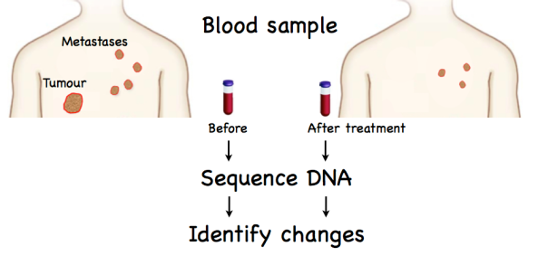
Curious behaviour though it may be, there’s nothing new about the idea of cells shedding bits of their genetic code – that was first shown to happen over 60 years ago. What is novel is the evidence that not only does this happen in a variety of cancer cells but that modern methods enable those fragments to be isolated from just a teaspoonful of blood: the sequence of the DNA can then be determined – which gives the mutational signature of the original tumour. A remarkable development has now shown that repeating these steps over a period of time can reveal the response of secondary tumours (metastases) to drug treatment (chemotherapy).

Untitled

One great advantage of this blood sampling method is that it is as near as makes no difference ‘non-invasive’. That is, it uses only a (small) blood sample and there’s no need for painful excavations to dig out tumour samples. The study, largely funded by Cancer Research UK, looked at three major cancers (breast, ovarian and lung) and identified specific mutations caused by drugs over a period of one to two years. For good measure they also took tumour samples to show that the mutation patterns found in circulating DNA did indeed represent what had gone on in the tumour itself. In other words, they had established what scientists like to call ‘proof of principle’ – i.e. we can do it!

There’s another more subtle advantage of this approach in that it gets round a problem we described in Molecular Mosaics: tumours are a mixture and the mutational signature differs depending on which bit you sample and sequence. The cell-free DNA fragments collected from blood are a gemisch – an averaged signature if you like – that may therefore give a better picture of the target for drug cocktails at any given time during tumour evolution.

Why is this so important?

There are two main reasons why it’s difficult to exaggerate the potential important of this step. The first is that metastasis accounts for over 90% of cancer deaths, the second that the fiendish ingenuity with which tumours negate chemotherapy, i.e. develop drug resistance, is one of the biggest challenges to successful treatment. So, the sooner changes that enable tumours to become insensitive to drugs can be detected the better in terms of adjusting the treatment regime. Even more exciting, however, is that notion that the DNA shed by cancers into the circulation may permit detection years or even decades earlier than is possible with any of the current methods (e.g., mammography) – with screening being carried put routinely from blood samples. Being even more optimistic, very early stage tumours may be particularly susceptible to appropriate drug combos, so that we might look forward to the day when chemotherapy replaces surgery as the first line of treatment for most cancers.

Reference

Murtaza, M. et al., (2013). Non-invasive analysis of acquired resistance to cancer therapy by sequencing of plasma DNA. Nature 497, 108–112.

Beware of Greeks …

Finding the words

One of the pitfalls of writing is repetition. Be it book, blog or broadsheet, most authors must dread someone gleefully chirruping ‘You used that exact phrase in 1999.’ I wonder if The Immortal Bard suffered likewise – having to resort to ferreting through piles of dusty manuscripts, finally in desperation shouting ‘Anne, Anne – got this great new line If you prick us do we not bleed? – Heard it before?’

‘Yes, of course dear. You used it in that thing about the Italian moneylender.’

‘Damn. Thought it sounded familiar. Where would I be without your memory – make me immortal with a kiss.’

‘Give over you daft beggar – even you know that’s one of that Marlowe bloke’s lines!’

‘Doh!’

I’m something of a sitting duck here, partly through not being Shakespeare but also because of the habit of often talking about biology. Take one simple example, Will’s tiny pinprick of blood – in which there will be about fifty million cells. That’s fifty million separate little sacs swirling around in a bead you wouldn’t notice if it wasn’t bright red. Isn’t that stunning for starters? Indeed, but it’s when we turn to molecular cancer that nature’s capacity to amaze is unfettered, the remarkable becomes the norm and even the English language can seem inadequate.

Finding the exit

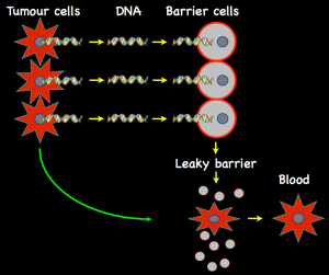
A study of a mouse model of one form of leukemia is the most recent contribution to have us sifting Shakespeare’s superlatives to do justice to the discovery. Blood cells start life within the bone marrow but, until they’ve matured, they’re corralled by a marrow–circulation barrier, also made of cells. Adult cells normally make proteins on their surface that attach to the barrier, and these help them to squeeze past into the freedom of the circulation. In leukemia abnormal levels of white blood cells are present in the circulation, which means that those cells have also found a way through the bone barrier. A Prague group have shown that one type of leukemic cell has come up with an astonishingly novel escape mechanism in which they release fragments of their own DNA. That’s pretty staggering because not only is DNA generally locked in the nucleus but it comes in large chunks called chromosomes. So two very unusual things have happened to get to this stage: (1) some of DNA has been shattered and (2) these pieces have crossed not only the membrane that encloses the nucleus but also the outer boundary of the cell itself.

DNA fragments from tumour cells enter barrier cells and kill them, releasing tumour cells into the circulation

DNA fragments from tumour cells enter barrier cells and kill them, releasing tumour cells into the circulation

But then something even more extraordinary happens: having tunneled their way out of the tumour cell, the escaped bits of DNA do a kind of reverse reprise by entering the cells that form the barrier between bone marrow and circulating blood. It’s as though the barrier cells see the passing packages of DNA as presents and gobble them up. Alas! They should have read their Virgil – or at least Dryden’s summary of the tale of the wooden horse of Troy: ‘Trust not their presents, nor admit the horse’ – for the barrier cells pay the ultimate price for their gluttony. The DNA fragments are sensed as something abnormal – as indeed they are – and this provokes a stress response – and a pretty extreme one at that – because the cells are so overwhelmed by the influx that they commit suicide.

The capacity for individual cells to switch on a death program is an important part of life – it’s essential in normal development and it’s also the best cancer defence we have. In other words, if things get out of control, kill the cell – because that eliminates the danger and cells can be replaced. But here we have an almost stupefying paradox: in the tumour cells this defence is neutralized – but they’ve come up with a way of turning it on in the normal cells they have to get past in order to spread around the organism.

It’s another astounding example of the plasticity of our genetic material and the incredible adaptability of cancer cells. Even Mr. S. might feel adjectivally challenged!

Reference

Dvořáková, M. et al., (2012). DNA released by leukemic cells contributes to the disruption of the bone marrow microenvironment. Oncogene 10 December 2012; doi: 10.1038/onc.2012.553

A Ray of Sunshine

One of the fascinating things about cancer is that it touches every aspect of biology. Of course, most will know that it’s caused by mutations – changes in the material that carries our genetic code. But many influences play on the genetic keyboard of DNA and those that are part of the world around us are a very mixed bunch. In Betrayed by Nature I split them into two: those we can do something about and the rest. The latter includes radiation from the ground… it’s all around us, we’ve evolved bathed in it and, apart from not living where the levels of radon are particularly high, there’s nothing we can do about it – so just forget it.

At the other end of the spectrum, so to speak, comes sunshine. We’ve evolved with that too – indeed we wouldn’t be here without it. Aside from driving photosynthesis in plants, humans use the radiant energy of the sun to make vitamin D (sometimes called the “sunshine vitamin”). Vitamin D deficiency is one cause of the childhood bone defect rickets, a condition that has reappeared in the UK in recent years because some kids are seeing less of the sun. So for humans catching the rays is desirable but we teeter along a sunny tightrope between what we need and what may ultimately be fatal. The risk comes from the ultra violet component of sunlight – radiation that has sufficient energy to damage DNA directly, making it a mutagen that can cause cancer. The cancer in question is, of course, melanoma that develops from abnormal moles on the skin. The global incidence of melanoma is increasing and, in the UK, about 90% of cases are estimated to be linked to exposure to ultra violet light. To most folk this means sunshine but those so inclined can walk the tightrope horizontally by using sunbeds (incredibly, in 1999 Cancer Research UK found that a quarter of men and a third of women questioned said they’d used a gizmo of this sort in the previous six months).

Which goes to show that human beings seem unable to resist the pursuit of the unattainable. The fair skinned think it cool to be darker whilst pharmaceutical giants are apparently making pots from selling creams to Indian ladies on the pitch that they will lighten their skin!

… and not so good

Good rays …

With a sigh for humanity let us pass from risks we take for no reason other than vanity or stupidity to those we may feel obliged to take as the lesser of two awkward options. There’s almost no chance that anyone reading this hasn’t had an X-ray of some sort. We have them to give our dentist a precise guide to the cause of our agony, rather than have him solve the problem by a series of trial and error excavations, or to tell our orthopaedic surgeon how best to go about piecing together the results of our latest stress-test on the human frame. We know X-rays are bad for us – they’re even more energetic than ultra violet radiation, so they’re a super-mutagen. Waves of cancer you might say.

So the issue here is one of choice. It’s a bit like a general anaesthetic: they do tend to make you throw up and about one in every 100,000 is fatal but, confronted with surgery, which would you vote for: a whiff of halothane or the offer of a slug of whisky and a rag to bite on? Computed tomography (CT) is an alternative application of X-rays but, instead of a single shot giving a two-dimensional image, CT acquires a large number of such images, taken as the radiation beam moves through the body, to give a 3-D picture. This can represent whole organs, and it has become an immensely powerful diagnostic tool since its introduction in the early 1970s. However, there’s no advance without anguish, and the additional information provided by a CT scan requires much more radiation than a traditional X-ray (typically 10 millisievert (mSv) compared with about 0.04 mSv for a chest X-ray). As our annual dose of “unavoidable” natural radiation is about 3 mSv it’s probably safe to say that these medical exposures are not a serious hazard – although babies in the womb are particularly sensitive to radiation. Even so, there are estimates that about 1% of USA cancers are due to CT scans, although there is no evidence that doses below 100 mSv induce tumours in animals.

A new study has enlarged the picture by finding that CT scans of children under 15 may increase the risk of leukemia and brain cancer. Three-fold increases were estimated for acute lymphoblastic leukaemia as a result of five to ten scans and for brain tumours by two or three scans. This sounds somewhat scary but it’s worth noting that these diseases are very rare in children. In the UK the incidence in under-20 year olds is just over four per 100,000 of leukemia – slightly less for brain or central nervous system cancers.

So the evidence indicates a small increase in an already low level of risk. As ever in life, therefore, it’s a matter of balance. The sensible advice for children (and everyone else for that matter) is not to have CT scans unless they are likely to provide critical clinical information that cannot be obtained by other means, for example, ultrasound or conventional X-rays.

Reference

Pearce, M.S., Salotti, J.A., Little, M.P., McHugh, K., Lee, C., Kim, K.P., Howe, N.L., Ronckers, C.M., Rajaraman, P., Craft, A.W., Parker, L. and de González, A.B. (2012). Radiation exposure from CT scans in childhood and subsequent risk of leukaemia and brain tumours: a retrospective cohort study. The Lancet 380, 499–505.